



Vol. 14. Núm. 12.
Páginas 641-650 (Junio 2024)

Actualización
Ascitis en el paciente no cirrótico

Ascites in non-cirrhotic patients

B. Burgueño GómezaI. Ruiz NúñezaM.E. Villacastín RuizbR. Pintado Garridob
Doi : https://10.1016/j.med.2024.06.001
Opciones
Resumen

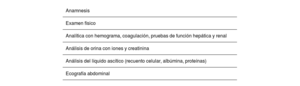
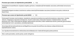
La ascitis, definida como la acumulación anormal de líquido en la cavidad peritoneal, es debida principalmente a cirrosis e hipertensión portal (HTP); sin embargo, en un 20% de los casos no se relaciona con dicha entidad. Su estudio es esencial para determinar el manejo terapéutico debido a su diversa etiología. La fisiopatología de la ascitis no cirrótica no está habitualmente relacionada con la HTP asociada a la cirrosis, y la comprensión de estos mecanismos facilita el diagnóstico etiológico. Los síntomas son: distensión abdominal, aumento de peso, plenitud postprandial e incluso disnea. La exploración física puede ser normal en casos de ascitis muy ligera, o bien identificar un aumento del perímetro abdominal en casos más avanzados o graves, también pueden aparecer signos asociados a la enfermedad subyacente. La clasificación según el Club Internacional de la Ascitis la divide en función de la cantidad y distensión abdominal. La evaluación inicial incluye: historia clínica, examen físico, ecografía abdominal y análisis de líquido ascítico. La paracentesis diagnóstica es crucial para identificar la causa, analizando características del líquido como su apariencia y contenido, especialmente la concentración de proteínas y el gradiente de albúmina sero-ascítico que permitirán discriminar las diferentes causas. En los países occidentales, las causas más frecuentes son la patología tumoral y la cardíaca, mientras que en los países en desarrollo es la patología infecciosa, sobre todo la tuberculosis, la más prevalente.

Palabras clave
Clasificación ascitis
Paracentesis
Cáncer
Diuréticos
Abstract

Ascites, defined as the abnormal accumulation of fluid in the peritoneal cavity, is mainly due to cirrhosis and portal hypertension (PHT). However, in 20% of cases, it is not related to these diseases. Given its varied etiology, it is essential to study it in order to determine therapeutic management. The pathophysiology of non-cirrhotic ascites is not usually related to cirrhosis-associated PHT, and understanding these mechanisms facilitates the etiologic diagnosis. Symptoms include abdominal distension, weight gain, postprandial fullness, and even dyspnea. The physical examination may be normal in cases of very mild ascites. An increase in abdominal circumference may be identified in more advanced or severe cases and signs associated with the underlying disease may also appear. The International Club of Ascites classification categorizes it according to the amount and abdominal distension. The initial evaluation includes a medical history, physical examination, abdominal ultrasound, and ascitic fluid analysis. Diagnostic paracentesis is crucial for identifying the cause, analyzing characteristics of the fluid such as its appearance and content, especially the protein concentration and the serum ascites albumin gradient, which will allow differentiating the different causes. In Western countries, the most frequent causes are tumor and heart diseases while in developing countries, infectious diseases, especially tuberculosis, are the most prevalent.

Keywords
Ascites classification
Paracentesis
Cancer
Diuretics

Identifíquese

¿Aún no es suscriptor de la revista?
Comprar el acceso al artículo

Comprando el artículo el pdf del mismo podrá ser descargado

Teléfono para incidencias

De lunes a viernes de 9h a 18h (GMT+1) excepto los meses de julio y agosto que será de 9 a 15h
Llamadas desde España 932 415 960
Llamadas desde fuera de España +34 932 415 960
Bibliografía
[1]
B.A. Runyon
202406250456584918
202406250456584919
.
Ascites and spontaneous bacterial peritonitis.
Sleisenger and Fordtran's gastrointestinal and liver diseases, 10ª ed, Elsevier, pp. 1517-1555
[2]
Santos
2024062504565849110
Santamaría L, García-Pozuelo Adalia N. Ascitis. Manual de diagnóstico y terapéutica médica. 9ª ed. Madrid: Hospital universitario 12 de Octubre. p. 891-900.
[3]
European
2024062504565849111
2024062504565849112
Association for the Study of the Liver.
EASL clinical practice guidelines on the management of ascites, spontaneous bacterial peritonitis, and hepatorenal syndrome in cirrhosis.
J Hepatol., (2010), 53 pp. 397-417
[4]
V. Arroyo
2024062504565849113
, M. Navasa.
Ascites and spontaneous bacterial peritonitis.
Schiff's diseases of the liver, 11th ed., John Wiley & Sons Ltd, pp. 527-567
[5]
L.J. Webb
2024062504565849114
, S. Sherlock.
The etiology, presentation and natural history of extra-hepatic portal venous obstruction.
Q J Med., (1979), 48 pp. 627
[6]
P. Carrier
2024062504565849115
2024062504565849116
, J. Jacques, M. Debette-Gratien, R. Legros, M. Sarabi, E. Vidal, et al.
L’ascite non liée à la cirrhose: physiopathologie, diagnostic et étiologies [Non-cirrhotic ascites: pathophysiology, diagnosis and etiology].
Rev Med Interne., (2014), 35 pp. 365-371
[7]
S.W. Biggins
2024062504565849117
, P. Angeli, G. Garcia-Tsao, P. Ginès, S.C. Ling, M.K. Nadim, et al.
Diagnosis, evaluation, and management of ascites, spontaneous bacterial peritonitis and hepatorenal syndrome: 2021 Practice Guidance by the American Association for the Study of Liver Diseases.
Hepatology., (2021), 74 pp. 1014-1048
[8]
S. Jang
2024062504565849118
, T. Stevens, R. Lopez, A. Bhatt, J.J. Vargo.
Superiority of gastrojejunostomy over endoscopic stenting for palliation of malignant gastric outlet obstruction.
Clin Gastroenterol Hepatol., (2019), 17
[9]
J.R. Mackey
2024062504565849119
, P.M. Venner.
Malignant ascites: demographics, therapeutic efficacy and predictors of survival.
Can J Oncol., (1996), 6 pp. 474-480
[10]
D.G. Jayne
2024062504565849120
, S. Fook, C. Loi, F. Seow-Choen.
Peritoneal carcinomatosis from colorectal cancer.
Br J Surg., (2002), 89 pp. 1545-1550
[11]
C.Y. Li
2024062504565849121
, T. Kennedy, H.R. Alexander.
Treatment of patients with malignant peritoneal mesothelioma.
J Clin Med., (2022), 11 pp. 1891
[12]
A.N. Husain
2024062504565849122
, T.V. Colby, N.G. Ordóñez, T.C. Allen, R.L. Attanoos, M.B. Beasley, et al.
Guidelines for pathologic diagnosis of malignant mesothelioma 2017 update of the consensus statement from the International Mesothelioma Interest Group.
Arch Pathol Lab Med., (2018), 142 pp. 89-108
[13]
P. Baas
2024062504565849123
, A. Scherpereel, A.K. Nowak, N. Fujimoto, S. Peters, A.S. Tsao, et al.
First-line nivolumab plus ipilimumab in unresectable malignant pleural mesothelioma (CheckMate 743): a multicentre, randomised, open-label, phase 3 trial.
Lancet., (2021), 397 pp. 375-386
[14]
N.J. Carr
2024062504565849124
, T.D. Cecil, F. Mohamed, L.H. Sobin, P.H. Sugarbaker, S. González-Moreno, et al.
Peritoneal Surface Oncology Group International. A consensus for classification and pathologic reporting of pseudomyxoma peritonei and associated appendiceal neoplasia: The results of the Peritoneal Surface Oncology Group International (PSOGI) modified delphi process.
Am J Surg Pathol., (2016), 40 pp. 14-26
[15]
N.J. Carr
2024062504565849125
, F. Bibeau, R.F. Bradley, P. Dartigues, R.M. Feakins, K.R. Geisinger, et al.
The histopathological classification, diagnosis and differential diagnosis of mucinous appendiceal neoplasms, appendiceal adenocarcinomas and pseudomyxoma peritonei.
Histopathology., (2017), 71 pp. 847-858
[16]
N.J. Carr
2024062504565849126
, F. Bibeau, R.F. Bradley, P. Dartigues, R.M. Feakins, K.R. Geisinger, et al.
The histopathological classification, diagnosis and differential diagnosis of mucinous appendiceal neoplasms, appendiceal adenocarcinomas and pseudomyxoma peritonei.
Histopathology., (2017), 71 pp. 847-858
[17]
H. Eraksoy
2024062504565849127
.
Gastrointestinal and abdominal tuberculosis.
Gastroenterol Clin North Am., (2021), 50 pp. 341-360
[18]
U. Vaid
2024062504565849128
, G.C. Kane.
Tuberculous peritonitis.
Microbiol Spectr., (2017), 5
[19]
A. Riquelme
2024062504565849129
, M. Calvo, F. Salech, S. Valderrama, A. Pattillo, M. Arellano, et al.
Value of adenosine deaminase (ADA) in ascitic fluid for the diagnosis of tuberculous peritonitis: a meta-analysis.
J Clin Gastroenterol., (2006), 40 pp. 705-710
[20]
A. Aamar
2024062504565849130
, K. Madhani, M.S. Anwar, P. Singh, J. Garsten.
Whipple's disease manifested as recurrent ascites.
Cureus., (2017), 9 pp. e1108
[21]
H.M. Shih
2024062504565849131
, M.J. Bair, H.L. Chen, I.T. Lin.
Eosinophilic gastroenteritis: Brief review.
Acta Gastroenterol Belg., (2016), 79 pp. 239-244
[22]
M.H. Su
2024062504565849132
, S.W. Cho, Y.S. Kung, J.H. Lin, W.L. Lee, P.H. Wang.
Update on the differential diagnosis of gynecologic organ-related diseases in women presenting with ascites.
Taiwan J Obstet Gynecol., (2019), 58 pp. 587-591
[23]
T. Gungor
2024062504565849133
, M. Kanat-Pektas, M. Ozat, M. Zayifoglu Karaca.
A systematic review: endometriosis presenting with ascites.
Arch Gynecol Obstet., (2011), 283 pp. 513-518
[24]
Practice
2024062504565849134
Committee of the American Society for Reproductive Medicine.
Electronic address: ASRM@asrm.org; Practice Committee of the American Society for Reproductive Medicine. Prevention and treatment of moderate and severe ovarian hyperstimulation syndrome: a guideline.
Fertil Steril., (2016), 106 pp. 1634-1647
[25]
A. Takahashi
2024062504565849135
, J. Takahama, N. Marugami, M. Takewa, T. Itoh, S. Kitano, et al.
Ectopic pregnancy: MRI findings and clinical utility.
Abdom Imaging., (2013), 38 pp. 844-850
[26]
R. Bhardwaj
2024062504565849136
, H. Vaziri, A. Gautam, E. Ballesteros, D. Karimeddini, G.Y. Wu.
Chylous ascites: A review of pathogenesis, diagnosis and treatment.
J Clin Transl Hepatol., (2018), 6 pp. 105-113