
Vol. 11. Núm. 28.
Páginas 1720-1727 (Marzo 2013)

Células presentadoras de antígeno

Antigen presenting cells

J. Monserrat SanzM. MartínE. ReyesA. Prieto Martín
Doi : https://10.1016/S0304-5412(13)70548-1
Opciones
Resumen

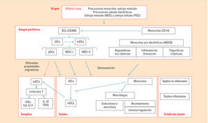
Las células presentadoras de antígeno profesionales (APC) representan un conjunto celular, perteneciente a nuestro sistema inmune innato, especializado en fagocitar o pinocitar microorganismos, hidrolizarlos en pequeños fragmentos moleculares e incorporarlos a sus correspondientes moléculas presentadoras de antígenos. Este conjunto de APC está compuesto principalmente por precursores en la médula ósea, monocitos circulantes en la sangre periférica, macrófagos en los tejidos, linfocitos B, células dendríticas y células de Langerhans en prácticamente todos los tejidos de nuestro organismo. Cada uno de estos tipos celulares está especializado en capturar, procesar y presentar antígenos de diferente naturaleza. Mediante moléculas de histocompatibilidad de clase II son capaces de presentar pequeños fragmentos peptídicos a los linfocitos T convencionales, mientras que mediante moléculas presentadoras como el CD1 son capaces de presentar pequeños fragmentos lipídicos a los linfocitos NKT. En este artículo nos vamos a centrar en el estudio de los monocitos, macrófagos y células dendríticas, y el avance que se ha experimentado en los últimos años en el estudio de las subpoblaciones que los componen. Asimismo, abordaremos el relevante papel de los monocitos en la inflamación y en la destrucción de patógenos y el papel de las células dendríticas como nexo entre la inmunidad innata y la adaptativa, encargándose de iniciar la activación de los linfocitos T novatos.

Palabras clave
Monocitos
Células dendríticas
Fagocitosis
Procesamiento antigénico
Moléculas de histocompatibilidad de clase II
CD1
Inflamación
Presentación antigénica
Abstract

Professional “antigen presenting cells” represent a cellular group, belonging to our innate immune system, specialized in phagocytizing or pinocytizing microorganisms, hydrolyzing them into small molecular fragments and incorporating them into their corresponding antigen presenting molecules. This combination of APC is principally made up of precursors in the bone marrow, peripheral circulating blood monocytes, macrophages in the tissues, B cells, dendritic cells and Langerhans cells in practically all the tissues in our body. Each one of these types of cells is specialized in capturing, processing and presenting different types of antigens. By class II histocompatibility molecules, they are capable of presenting small peptidic fragments to the conventional T cells, while by presenting molecules such as the CD1, they are capable of presenting small lipidic fragments to the NKT lymphocytes. In this chapter, we are going to focus on the study of the monocytes, macrophages and dendritic cells and the advance experienced in recent years in the study of the subpopulations they make up. We will also address the relevant role of monocytes in inflammation and the destruction of pathogens and the role of dendritic cells as a link between the adaptive and innate immunity, taking charge of initiating the activation of the novel T cells.

Keywords
Monocytes
Dendritic cells
Phagocytosis
Antigenic processing
Class II histocompatibility molecules
CD1
Inflammation
Presentation

Identifíquese

¿Aún no es suscriptor de la revista?

Comprar el acceso al artículo

Comprando el artículo el pdf del mismo podrá ser descargado

Teléfono para incidencias

De lunes a viernes de 9h a 18h (GMT+1) excepto los meses de julio y agosto que será de 9 a 15h

Llamadas desde España 932 415 960
Llamadas desde fuera de España +34 932 415 960
Bibliografía
[1.]
C. Auffray, M.H. Sieweke, F. Geissmann.
Blood monocytes: development, heterogeneity, and relationship with dendritic cells.
Annu Rev Immunol, (2009), 27 pp. 669-692
[2.]
C. Auffray, D.K. Fogg, E. Narni-Mancinelli, B. Senechal, C. Trouillet, N. Saederup, et al.
CX3CR1+ CD115+ CD135+ common macrophage/DC precursors and the role of CX3CR1 in their response to inflammation.
J Exp Med, (2009), 206 pp. 595-606
[3.]
D.A. Hume.
Differentiation and heterogeneity in the mononuclear phagocyte system.
Mucosal Immunol., (2008), 1 pp. 432-441
[4.]
F. Geissmann, S. Jung, D.R. Littman.
Blood monocytes consist of two principal subsets with distinct migratory properties.
Immunity., (2003), 19 pp. 71-82
[5.]
P. Ancuta, L. Weiss, N. Haeffner-Cavaillon.
CD14+CD16++ cells derived in vitro from peripheral blood monocytes exhibit phenotypic and functional dendritic cell-like characteristics.
[6.]
H. Jahnisch, S. Fussel, A. Kiessling, R. Wehner, S. Zastrow, M. Bachmann, et al.
Dendritic cell-based immunotherapy for prostate cancer.
Clin Dev Immunol, (2010), 2010 pp. 517493
[7.]
W. Xu, X. Zhao, M.R. Daha, K.C. van.
Reversible differentiation of pro- and anti-inflammatory macrophages.
Mol Immunol, (2013), 53 pp. 179-186
[8.]
D. Hasan, N. Chalouhi, P. Jabbour, T. Hashimoto.
Macrophage imbalance (M1 vs M2) and upregulation of mast cells in wall of ruptured human cerebral aneurysms: preliminary results.
J Neuroinflammation, (2012), 9 pp. 222
[9.]
S. Gordon.
Macrophag es and phagocytosis.
Fundamental immunology, 6ª, Lippincott Williams and Wilkins, pp. 547
[10.]
F. Geissmann, C. Auffray, R. Palframan, C. Wirrig, A. Ciocca, L. Campisi, et al.
Blood monocytes: distinct subsets, how they relate to dendritic cells, and their possible roles in the regulation of T-cell responses.
Immunol Cell Biol., (2008), 86 pp. 398-408
[11.]
J. Skrzeczynska-Moncznik, M. Bzowska, S. Loseke, E. Grage-Griebenow, M. Zembala, J. Pryjma.
Peripheral blood CD14high CD16+ monocytes are main producers of IL-10.
Scand J Immunol., (2008), 67 pp. 152-159
[12.]
H.W. Ziegler-Heitbrock.
Definition of human blood monocytes.
J Leukoc Biol., (2000), 67 pp. 603-606
[13.]
G. Fingerle-Rowson, J. Auers, E. Kreuzer, P. Fraunberger, M. Blumenstein, L.H. Ziegler-Heitbrock.
Expansion of CD14+CD16+ monocytes in critically ill cardiac surgery patients.
Inflammation., (1998), 22 pp. 367-379
[14.]
A. Horelt, K.U. Belge, B. Steppich, J. Prinz, L. Ziegler-Heitbrock.
The CD14+CD16+ monocytes in erysipelas are expanded and show reduced cytokine production.
[15.]
K. Mizuno, T. Toma, H. Tsukiji, H. Okamoto, H. Yamazaki, K. Ohta, et al.
Selective expansion of CD16high.
Clin Exp Immunol, (2005), 142 pp. 461-470
[16.]
F. Tacke, G.J. Randolph.
Migratory fate and differentiation of blood monocyte subsets.
Immunobiology., (2006), 211 pp. 609-618
[17.]
M. Siedlar, M. Frankenberger, L.H. Ziegler-Heitbrock, K.U. Belge.
The M-DC8-positive leukocytes are a subpopulation of the CD14+ CD16+ monocytes.
Immunobiology., (2000), 202 pp. 11-17
[18.]
R. Thomas, P.E. Lipsky, subsets. Human peripheral blood dendritic cell.
Isolation and characterization of precursor and mature antigen-presenting cells.
J Immunol., (1994), 153 pp. 4016-4028
[19.]
J. Almeida, C. Bueno, M.C. Alguero, M.L. Sánchez, M. de Santiago, L. Escribano, et al.
Comparative analysis of the morphological, cytochemical, immunophenotypical, and functional characteristics of normal human peripheral blood lineage(–)/CD16(+)/HLA-DR(+)/CD14(–/lo) cells, CD14(+) monocytes, and CD16(-) dendritic cells.
Clin Immunol., (2001), 100 pp. 325-338
[20.]
Q. Huang, Y. Ma, A. Adebayo, R.M. Pope.
Increased macrophage activation mediated through toll-like receptors in rheumatoid arthritis.
Arthritis Rheum., (2007), 56 pp. 2192-2201
[21.]
C. Hespel, M. Moser.
Role of inflammatory dendritic cells in innate and adaptive immunity.
Eur J Immunol, (2012), 42 pp. 2535-2543
[22.]
G.J. Randolph, S. Beaulieu, S. Lebecque, R.M. Steinman, W.A. Muller.
Differentiation of monocytes into dendritic cells in a model of transendothelial trafficking.
Science, (1998), 282 pp. 480-483
[23.]
L. Castiello, M. Sabatino, P. Jin, C. Clayberger, F.M. Marincola, A.M. Krensky, et al.
Monocyte-derived DC maturation strategies and related pathways: a transcriptional view.
Cancer Immunol Immunother, (2011), 60 pp. 457-466
[24.]
M. Moser.
Dendritic cells.
Fundamental immunology, 6ª, Lippincott Williams and Wilkins, pp. 451
[25.]
S. Jaitley, T. Saraswathi.
Pathophysiology of Langerhans cells.
J Oral Maxillofac Pathol, (2012), 16 pp. 239-244
[26.]
R.M. Steinman, Z.A. Cohn.
Identification of a novel cell type in peripheral lymphoid organs of mice.I. Morphology, quantitation, tissue distribution.
J Exp Med, (1973), 137 pp. 1142-1162
[27.]
T. Zhou, Y. Chen, L. Hao, Y. Zhang.
DC-SIGN and immunoregulation.
Cell Mol Immunol, (2006), 3 pp. 279-283
[28.]
N.K. van, G.C. de.
Human follicular dendritic cells: function, origin and development.
Semin Immunol, (2002), 14 pp. 251-257
[29.]
A. Dzionek, A. Fuchs, P. Schmidt, S. Cremer, M. Zysk, S. Miltenyi, et al.
BDCA-2BDCA-3, and BDCA-4: three markers for distinct subsets of dendritic cells in human peripheral blood.
J Immunol, (2000), 165 pp. 6037-6046
[30.]
D.H. Margulies, K. Natarajan, J. Rossjohn, J. McCluskey.
Major histocompatibility complex molecules: structure, function and genetics.
Fundamental immunology, Sixth edition, Lippincott Williams and Wilkins, pp. 570
Copyright © 2013. Elsevier España, S.L.. Todos los derechos reservados