

Vol. 14. Núm. 7.
Páginas 402-407 (Abril 2024)

Protocolos de práctica asistencial
Protocolo diagnóstico y de seguimiento de las lesiones quísticas pancreáticas

Diagnostic and follow-up protocol for pancreatic cystic lesions

V. Flores-FernándezA. Hernando AlonsoL. Pérez-Carazo
Doi : https://10.1016/j.med.2024.04.010
Opciones
Resumen

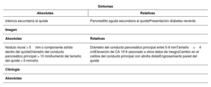
Las lesiones quísticas pancreáticas suelen ser un hallazgo incidental al realizar un estudio de imagen por otros motivos. El origen de estos quistes es variado y, según este, pueden ser benignas o tener potencial de malignidad; sin embargo, diferenciarlas no siempre es sencillo. Disponemos de varias herramientas para intentar identificar a qué tipo de quiste nos estamos enfrentando: ecoendoscopia, resonancia magnética, biomarcadores serológicos, quísticos e histológicos, pero no todos tienen una potencia diagnóstica óptima. A la hora de establecer el seguimiento, siempre debe tenerse en cuenta si el paciente tiene indicación quirúrgica; si es así, diferentes sociedades científicas han establecido estrategias de seguimiento y manejo de estas lesiones en función de la presencia de datos de riesgo de malignidad.

Palabras clave
Quiste pancreático
Neoplasia quística pancreática
Ecoendoscopia
Tumor mucinoso intraductal
Abstract

Pancreatic cystic lesions are usually an incidental finding when performing an imaging study for other reasons. The origin of these cysts varies and, depending on their origin, they can be benign or potentially malignant. However, differentiating between the two is not always easy. Several tools are available to try to identify the type of cyst—endoscopic ultrasound; magnetic resonance imaging; and serological, cystic, and histological biomarkers—but not all of them have optimal diagnostic power. When determining follow-up, whether the patient has an indication for surgery must always be taken into account. If so, various scientific societies have established strategies for the follow-up and management of these lesions according to the presence of signs of malignancy risk.

Keywords
Pancreatic cyst
Pancreatic cystic neoplasm
Endoscopic ultrasound
Intraductal mucinous tumor

Identifíquese

¿Aún no es suscriptor de la revista?
Comprar el acceso al artículo

Comprando el artículo el pdf del mismo podrá ser descargado

Teléfono para incidencias

De lunes a viernes de 9h a 18h (GMT+1) excepto los meses de julio y agosto que será de 9 a 15h
Llamadas desde España 932 415 960
Llamadas desde fuera de España +34 932 415 960
Bibliografía
[1]
European
202404150548456178
202404150548456179
Study Group on Cystic Tumours of the Pancreas.
European evidence-based guidelines on pancreatic cystic neoplasms.
[2]
M. Del Chiaro, R. Segersvärd, R. Pozzi Mucelli, E. Rangelova, N. Kartalis, C. Ansorge, et al.
Comparison of preoperative conference-based diagnosis with histology of cystic tumors of the pancreas.
Ann Surg Oncol., (2014), 21 pp. 1539-1544
[3]
J.M. Scheiman
2024041505484561710
2024041505484561711
, J.H. Hwang, P. Moayyedi.
American gastroenterological association technical review on the diagnosis and management of asymptomatic neoplastic pancreatic cysts.
Gastroenterology., (2015), 148 pp. 824-848
[4]
G.D. Thornton
2024041505484561712
, M.J. McPhail, S. Nayagam, M.J. Hewitt, P. Vlavianos, K.J. Monahan.
Endoscopic ultrasound guided fine needle aspiration for the diagnosis of pancreatic cystic neoplasms: a metaanalysis.
Pancreatology., (2013), 13 pp. 48-57
[5]
D.G. Ribaldone, M. Bruno, S. Gaia, A. Cantamessa, A. Bragoni, P. Caropreso, et al.
Differential diagnosis of pancreatic cysts: A prospective study on the role of intra-cystic glucose concentration.
Dig Liver Dis., (2020), 52 pp. 1026-1032
[6]
G.H. Elta
2024041505484561713
2024041505484561714
, B.K. Enestvedt, B.G. Sauer, A.M. Lennon.
ACG Clinical guideline: Diagnosis and management of pancreatic cysts.
Am J Gastroenterol., (2018), 113 pp. 464-479
[7]
D.R. Westerveld
2024041505484561715
, S.A. Ponniah, P.V. Draganov, D. Yang.
Diagnostic yield of EUS-guided through-the-needle microforceps biopsy versus EUS-FNA of pancreatic cystic lesions: a systematic review and meta-analysis.
Endosc Int Open., (2020), 8 pp. E656-E667
[8]
K. Kamata, M. Kitano, S. Omoto, K. Kadosaka, T. Miyata, K. Yamao, et al.
Contrast-enhanced harmonic endoscopic ultrasonography for differential diagnosis of pancreatic cysts.
Endoscopy., (2016), 48 pp. 35-41
[9]
R. Rosenblatt
2024041505484561716
, V. Dorfman, I. Epelboym, J.M. Poneros, A. Sethi, C. Lightdale, et al.
Demographic features and natural history of intermediate-risk multifocal versus unifocal intraductal papillary mucinous neoplasms.
Pancreas., (2015), 44 pp. 478-483
[10]
J.W. Park, J.Y. Jang, M.J. Kang, W. Kwon, Y.R. Chang, S.W. Kim.
Mucinous cystic neoplasm of the pancreas: Is surgical resection recommended for all surgically fit patients?.
Pancreatology., (2014), 14 pp. 131-136
[11]
N. Lubezky, M. Papoulas, Y. Lessing, G. Gitstein, E. Brazowski, I. Nachmany, et al.
Solid pseudopapillary neoplasm of the pancreas: Management and long-term outcome.
Eur J Surg Oncol., (2017), 43 pp. 1056-1060
[12]
J.K. Law, A. Ahmed, V.K. Singh, V.S. Akshintala, M.T. Olson, S.P. Raman, et al.
A systematic review of solid-pseudopapillary neoplasms: are these rare lesions?.
Pancreas., (2014), 43 pp. 331-337
[13]
K. Kwok
2024041505484561717
, J. Chang, L. Duan, B.Z. Huang, B.U. Wu.
Competing risks for mortality in patients with asymptomatic pancreatic cystic neoplasms: Implications for clinical management.
Am J Gastroenterol., (2017), 112 pp. 1330-1336