


Vol. 14. Núm. 7.
Páginas 392-396 (Abril 2024)

Protocolos de práctica asistencial
Protocolo diagnóstico de la colestasis

Diagnostic protocol for cholestasis

A. Díaz GómezI. Payeras OteroC. Iniesta CaveroV. Flores-Fernández
Doi : https://10.1016/j.med.2024.04.008
Opciones
Resumen

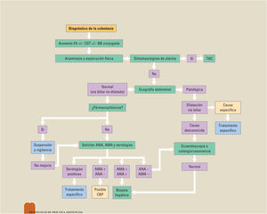
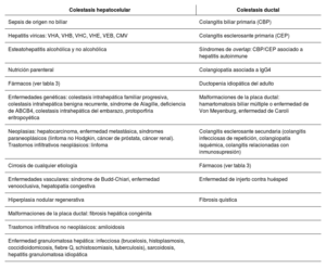
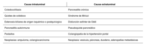
El concepto de colestasis hace referencia a la dificultad o detención del flujo biliar. Es un problema clínico frecuente que puede ser la manifestación de múltiples enfermedades. Se diferencia entre colestasis intrahepática cuando existe una alteración dentro del parénquima hepático, y colestasis extrahepática si la obstrucción se localiza a nivel de colédoco. La anamnesis y la exploración física minuciosa son imprescindibles para poder orientar el diagnóstico. La ecografía abdominal es la primera prueba de imagen a realizar. Ante una ecografía normal o no concluyente, se debe ampliar el estudio con otras pruebas de imagen más específicas. Si existe sospecha de malignidad, la tomografía computarizada abdominal debe ser el primer procedimiento diagnóstico. La ecoendoscopia oral y la colangiografía mediante resonancia magnética son exploraciones con una alta rentabilidad diagnóstica equiparable entre sí. La colangiopancreatografía retrógrada endoscópica permite una visualización directa de la vía biliar con la posibilidad de realizar terapéutica.

Palabras clave
Colestasis intrahepática
Colestasis extrahepática
Ecografía abdominal
Colangiografía por resonancia magnética
Abstract

The concept of cholestasis refers to difficulty or stoppage of bile flow. It is a common clinical problem that can be the manifestation of multiple diseases. A distinction can be drawn between intrahepatic cholestasis, when there is abnormality in the liver parenchyma, and extrahepatic cholestasis, if the obstruction is located in the common bile duct. A detailed case history and physical examination are essential in order to guide the diagnosis. An abdominal ultrasound is the first imaging test to be performed. In the presence of a normal or inconclusive ultrasound, the study should be expanded with other more specific imaging tests. If malignancy is suspected, an abdominal computed tomography scan should be the first diagnostic procedure. An oral endoscopic ultrasound and magnetic resonance cholangiopancreatography are examinations with a high diagnostic yield that is comparable between them. Endoscopic retrograde cholangiopancreatography allows for direct visualization of the bile duct with the possibility of performing treatment.

Keywords
Intrahepatic cholestasis
Extrahepatic cholestasis
Abdominal ultrasound
Magnetic resonance cholangiopancreatography

Identifíquese

¿Aún no es suscriptor de la revista?
Comprar el acceso al artículo

Comprando el artículo el pdf del mismo podrá ser descargado

Teléfono para incidencias

De lunes a viernes de 9h a 18h (GMT+1) excepto los meses de julio y agosto que será de 9 a 15h
Llamadas desde España 932 415 960
Llamadas desde fuera de España +34 932 415 960
Bibliografía
[1]
D.S. Pratt.
Liver chemistry and function tests.
Sleisenger y Fordtran's enfermedades digestivas y hepáticas, 10a ed, Elsevier, pp. 1243-1253
[2]
European
202404150548355938
202404150548355939
Association for the Study of the Liver.
EASL clinical practice guidelines: management of cholestatic liver diseases.
J Hepatol., (2009), 51 pp. 237-267
[3]
T. Álvarez-Nava Torrego.
Ictericia. En: Manual de diagnóstico y terapéutica médica.
8ª ed, MSD, (2016),
[4]
S. Saini
2024041505483559310
2024041505483559311
.
Imaging of the hepatobiliary tract.
N Engl J Med., (1997), 336 pp. 1889-1894
[5]
V.A. Sahni
2024041505483559312
2024041505483559313
, K.J. Mortele.
Magnetic resonance cholangiopancreatography: current use and future applications.
Clin Gastroenterol Hepatol., (2008), 6 pp. 967-977
[6]
ASGE
2024041505483559314
2024041505483559315
Standards of Practice Committee, J.L. Buxbaum, S.M. Abbas Fehmi, S. Sultan, D.S. Fishman, B.J. Qumseya, V.K. Cortessis, et al.
ASGE guideline on the role of endoscopy in the evaluation and management of choledocholithiasis.
Gastrointest Endosc., (2019), 89
[7]
M. Suzuki
2024041505483559316
, Y. Sekino, K. Hosono, K. Yamamoto, K. Kawana, H. Nagase, et al.
Endoscopic ultrasound versus magnetic resonance cholangiopancreatography for the diagnosis of computed tomography-negative common bile duct stone: Prospective randomized controlled trial.
Dig Endosc., (2022), 34 pp. 1052-1059
[8]
S. Afzalpurkar
2024041505483559317
2024041505483559318
, S. Giri, S. Kasturi, S. Ingawale, S. Sundaram.
Magnetic resonance cholangiopancreatography versus endoscopic ultrasound for diagnosis of choledocholithiasis: an updated systematic review and meta-analysis.
Surg Endosc., (2023), 37 pp. 2566-2573
[9]
L. Bianchi
2024041505483559319
.
Liver biopsy in elevated liver functions tests? An old question revisited.
J Hepatol., (2001), 35 pp. 290-294
[10]
I. Salueña
2024041505483559320
, L. Ortega, M.J. Devesa, G. López-Alonso, C. Taxonera, M. Díaz-Rubio, et al.
Utilidad de la biopsia hepática en el diagnóstico etiológico de las alteraciones de la analítica hepática de causa incierta [Utility of liver biopsy in the etiologic diagnosis of biochemical liver abnormalities of unknown cause].
Gastroenterol Hepatol., (2007), 30 pp. 325-330